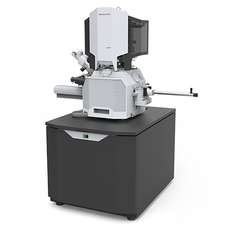
The Aquilos Cryo-FIB is a focused ion beam/scanning electron microscope system dedicated to imaging and preparation of thin lamella samples under cryogenic conditions.
Specifications/Capabilities
- Special sample holders for shallow-angle milling of EM grids
- Sample shuttle for autogrids: Cryo-FIB shuttle with integrated shutter system during cryo-transfers
- In-chamber retractable sputter coater for conductive coatings application
- GIS-system for protective coatings application
- MAPS Software for image correlation and lamella milling
- Non-immersion field emission-SEM column
- Everhart-Thornley SE Detector (ETD), segmented lower (T1) and upper (T2) detectors
- Schottky field emission gun
- FIB-Ga Ion Source
Applications
- Cryogenic imaging of beam-sensitive materials
- Cryo SEM and cryo-tomography
- Fabrication of lamellas for TEM imaging
Staff/Contact
Location
12-0144
MIT.nano (basement level)
60 Vassar Street (rear)
Cambridge, MA