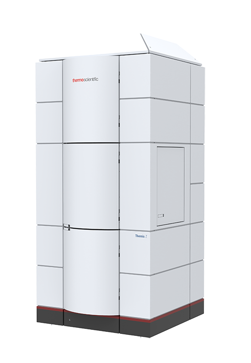
The Thermo Fisher Scientific (TFS) Themis Z G3 aberration-corrected scanning transmission electron microscope (STEM) achieves < 0.6 Å resolution (sample dependent) and is equipped with an optimized monochromator for < 30 meV energy resolution in EELS at 60 kV, a Gatan Continuum electron energy loss spectrometer and image filter, a TFS Super-X energy dispersive x-ray spectrometer system, a segmented STEM detector for fast DPC and iDPC imaging, a Lorentz lens, a 16 megapixel Ceta II camera and a 4D STEM EMPAD camera. Single tilt, double tilt, and tomography holders are available. The instrument can be operated at 60, 200, and 300 kV for the flexibility to accommodate a range electron beam sensitive materials.
- 60, 200 and 300 kV operating range
- Monochromator with < 30 meV energy resolution in EELS (vibrational spectroscopy and automated alignment software)
- Piezoelectric stage for compensating drift and fine sample movements
- Sub-1 Å imaging at 60 kV (particularly useful for 2D materials)
- Gatan Continuum spectrometer and imaging filter (atomic resolution chemical mapping with monochromated EELS)
- Super X-4 quadrant EDS detector with separable signals from each detector (atomic resolution elemental mapping with energy-dispersive X-ray spectroscopy (EDS))
- Imaging and EDS tomography to reconstruct structure and chemistry in 3D
- Thermo Fisher Scientific EMPAD detector
- Fast 4k x 4k CMOS camera
- Segmented diode detector for fast mapping of electromagnetic fields (differential phase contrast) and light elements (integrated differential phase contrast)
- Magnetic domains imaging via Lorentz lens settings
Accessory in-situ holders
- Mel-Build Atmos Double Tilt LN2 Vacuum/Inert Gas Transfer Holder
- DENS solutions Lightning Heating and Biasing Double Tilt Holder
- Hummingbird Liquid Flow Holder with electrochemistry
- Atomic-resolution imaging and spectroscopy
- Correlating morphology, structure, and chemistry
- Magnetic nanostructure characterization
- Tomography for imaging and 3D elemental Distributions
- Elemental and chemical analyses of small analytical volumes
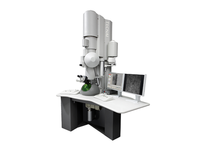
FEI Tecnai Multipurpose Digital TEM
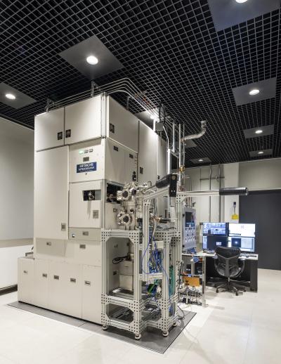
Hitachi HF 5000 Environmental S/TEM
Aubrey Penn, PhD
12-0178
Building 12 (basement)
60 Vassar Street
Cambridge, MA
Yong Zhang
12-0180
60 Vassar Street (rear)
Cambridge, MA
ASSISTED USE / TRAINING REQUEST
Prior electron microscopy imaging experience is required to become an independent user
12-0183
MIT.nano (basement level)
60 Vassar Street (rear)
Cambridge, MA