Label-free chemical imaging using the STELLARIS Coherent Raman Scattering (CRS) microscope enables you to obtain crucial biochemical and metabolic information that cannot be visualized with traditional fluorescent microscopy methods.
With STELLARIS CRS, you can image a wide range of specimens at the required high speed and resolution to follow essential processes of life. Take advantage of different modalities to maximize the information you get from your sample: Stimulated Raman Scattering (SRS), Coherent Anti-Stokes Raman Scattering (CARS), Second Harmonic Generation (SHG), two-photon fluorescence, and visible confocal fluorescence.
Capabilities include:
- Coherent Raman Imaging
- Coherent Raman Spectroscopy
- Label Free Imaging
- Chemical Mapping
Raman-Based Measurement Modalities
- Stimulated Raman Scattering (SRS) Imaging/Spectroscopy
- Coherent Anti-Stokes Raman Scattering (CARS) Imaging/Spectroscopy
Laser Excitation
- CRS Pump: 720 -980 nm (Tunable)
- CRS Stokes: 1032 nm (Fixed)
Spectral Range
- SRS: 507 – 3500 cm-1
- CARS: 1200 – 3500cm-1
Spectral Resolution: 1 cm-1 (Wavelength Dependent)
Spatial Resolution (lateral): < 500 nm (Wavelength and Objective Dependent)
Spatial Resolution (axial): < 2 um (Wavelength and Objective Dependent)
- Second Harmonic Generation (SHG) Imaging
- Two-Photon Fluorescence Imaging
- Visible Fluorescence Imaging
- Fluorescence Lifetime Imaging
Please make sure to acknowledge MIT.nano Characterization in any publication, presentations, and patents involving results originated from the use of the Leica Stellaris Confocal Raman at MIT.nano Characterization Focus Facilities or through assistance from MIT.nano staff.
Suggested language: "This work was performed in part in the MIT.nano Characterization Facilities"
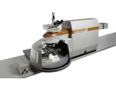
Renishaw Invia Reflex Micro Raman
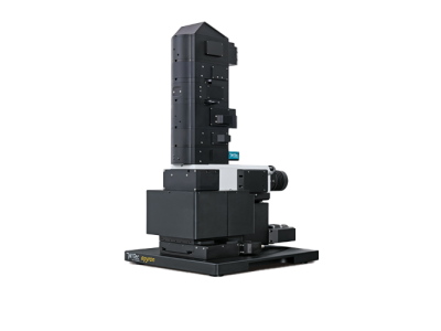
WITec alpha300 apyron Confocal Raman

12-0195B (EM12)
MIT.nano (basement level)
60 Vassar Street (rear)
Cambridge, MA